Each of the fundamental tissues is formed by several types of cells and typically by specific associations of cells and. The accuracy of cytology was higher in pure AIS 615 and GIL I II 222 than in mixed le-.

Typical Cytological Features Of Adenocarcinoma In Situ A Download Scientific Diagram
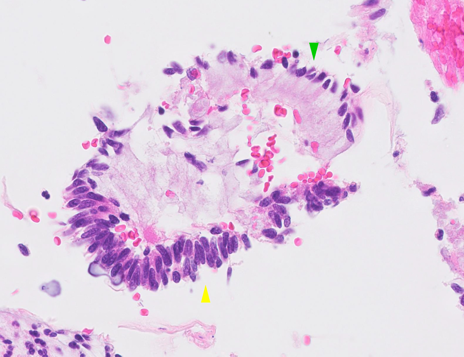
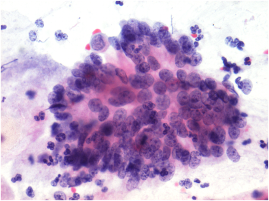
In contrast to the normal simple endocervical epithelium AIS shows pseudostratification of enlarged hyperchromatic nuclei numerous mitotic figures often apical and apoptosis.
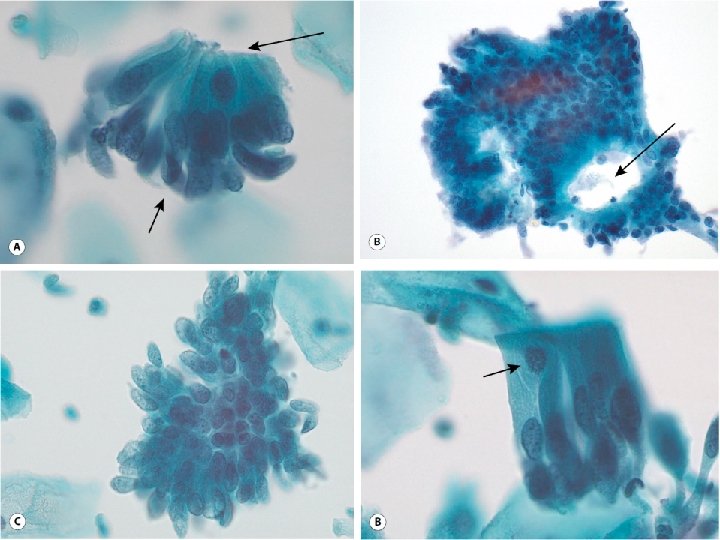
Image type of ais in pseudostratification cytology. Atypical endocervical cells most likely from a reparative process CP. Feathering is another distinctive feature of AIS. Endocervical cells with prominent pseudostratification with regular polarity of nuclei Endocervical Adenocarcinoma Intestinal type endocervical adenocarcinoma in situ Intestinal type endocervical adenocarcinoma 82 pathologists.
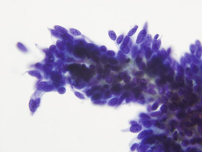
Endocervical type of AIS is characterized by moderate to high cellularity of tight clusters and cohesive sheets of glandular cells showing pseudostratification rosette formation palisades secondary gland openings and feathering at the edge of sheets. Find the perfect cytology stock photo. Gynaecological cytology in the form of Pap Papanicolaou smears is the most recognised type of test.
SIL in Glands Left image. If the context is clear it may be referred to as adenocarcinoma in situ abbreviated AIS. Specimens are obtained mechanically by scrapings or needle aspirates or by collection of exfoliated material from various body fluids.
Makes up 05 of all cervical cytology. However a wide variety of. Type of epithelial alteration 756 vs.
About 80 of cervical adenocarcinoma is of the usual type which is made up of medium sized mucin-poor glands with eosinophilic cytoplasm many mitoses and apoptotic debris The nuclei are hyperchromatic more elongate than round and have scattered mitotic figures Fig. Ais cytology cytology in outline format with mouse over histology previews. EC AC 70 pathologists.
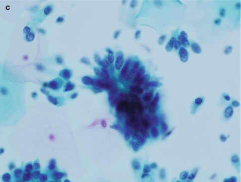
Cells in AIS clusters may arrange in a circular pattern with peripheral location of nuclei. 2E66Y XA7Z73 - other specified carcinoma in situ of cervix uteri cervical canal Epidemiology. AIS exhibits groups with a greater depth-of-focus and nuclear crowding with finely granular or vacuolated cytoplasm.
Huge collection amazing choice 100 million high quality affordable RF and RM images. 12 Interestingly patients with intestinal-type endocervical AIS are on average older 445 vs. Figure 46 a AIS in Pap smear Figure 46 b typical appearance of AIS on cytology.
Type of epithelial alteration 756 vs. AIS In AIS nucleoli may be present. Uncommon 1 of cervical noninvasive lesions versus 99 high grade squamous intraepithelial lesion HSIL in the SEER registry Mean age 38 years 10 - 15 years younger than invasive endocervical adenocarcinoma.
The accuracy of diagnosis of cell type obtained from sputum cytology bronchial aspirate bronchial biopsy or percutaneous lung biopsy in 161 cases of confirmed primary lung cancer has been examined and the pretreatment histological diagnosis has been compared with the final diagnosis made after surgical resection or necropsy. 553 and abnormalities of columnar epithelium 957. Affordable and search from millions of royalty free images photos and vectors.
Thousands of new high-quality pictures added every day. Endocervical-type adenocarcinoma is the commonest 70 of adenocarcinomas and may be preceded by adenocarcinoma in situ AIS also known as cervical glandular intraepithelial neoplasia CGIN which is a known precursor of adenocarcinoma Ostör et al. However polarity of cells in AIS is maintained.
AIS adenocarcinoma in situ. A glandular abnormality was detected in 32 of 72 AIS cases 444 and 37 of 48 ECCA cases 771. Cell biology encompasses both prokaryotic and eukaryotic cells and can be divided into many sub-topics which may include the study of cell metabolism cell communication cell cycle biochemistry and cell composition.
In terms of differential diagnosis cytology showed higher accu-racy in predicting lesion severity vs. Image from The Bethesda System for Reporting Cervical Cytology. 41aThe nuclear cytology is generally of intermediate grade.
Histology is the study of the tissues of the body and of how these tissues are arranged to constitute organs. Sectioning techniques cytology uses various cell preparation technologies. Sheet of cells that demonstrate nuclear enlargement increased nuclear to cytoplasmic NC ratios prominent sometimes multiple nucleoli and mitotic activity.
Endocervical adenocarcinoma in situ also adenocarcinoma in situ of the uterine endocervix is pre-invasive change of the uterine endocervixIt is closely tied to HPV infection. The intestinal-type AIS often stains less intensely with p16. Of the 120 patients with histologic AIS or ECCA 108 90 had abnormal Pap results including 66 of 72 92 with AIS and 42 of 48 875 with ECCA.
The positive rates of conventional sputum cytology and brush cytology were 16 and 32 which was lower than that of DNA aneuploidy detection by the automated image cytometry P005. Download Cytology stock photos. For the cytology see Endocervical adenocarcinoma in situ cytology.
No need to register buy now. Routine screen from a 39-year-old woman. Cell biology also cellular biology or cytology is a branch of biology that studies the structure function and behavior of cells.
In terms of differential diagnosis cytology showed higher accuracy in predicting lesion severity vs. In AIS the clusters of endocervical cells may also show pseudostratification with nuclei seen at different levels within the groups. 326 years and multiple studies have shown that the frequency of HPV detection in intestinal-type AIS is less than that for other forms of AIS with a corresponding lower rate of p16 diffuse positivity.
Find cytology stock images in HD and millions of other royalty-free stock photos illustrations and vectors in the Shutterstock collection. Nuclear elongation and overlap are common and correlate with the histological findings of nuclear pseudostratification and cellular crowding. The neoplastic endocervical cells of endocervical AIS and endocervical carcinoma Figure 21.
Histologic diagnosis after AGC on Cytology Adenocarcinoma In Situ AIS Squamous intraepithelial lesion any Most common pathology with AGC Often coexist with glandular lesions Adenocarcinoma Cervix endometrium tube ovary metastatic Reactive reparative polyps Microglandular hyperplasia from OCPs. AIS This small group of cells shows pseudostratification with crowded hyperchromatic nuclei. 553 and abnormalities of columnar epithe-lium 957.
Professor asastent of department histology cytology and embryology - KHODOROVSKA ALLA.
Pathology Outlines Adenocarcinoma In Situ
Cytology Of Glandular Lesions Basicmedical Key

Adenocarcinoma In Situ Ais Endocervical Glandular Epithelium Is Download Scientific Diagram
Cytopathology Basicmedical Key
Glandular Abnormalities Of The Cervix Eurocytology


Tidak ada komentar